


COVID-19 Update [August '22]
30 Aug 2022
By B. Jeanne Billioux, MD and Avindra Nath, MD
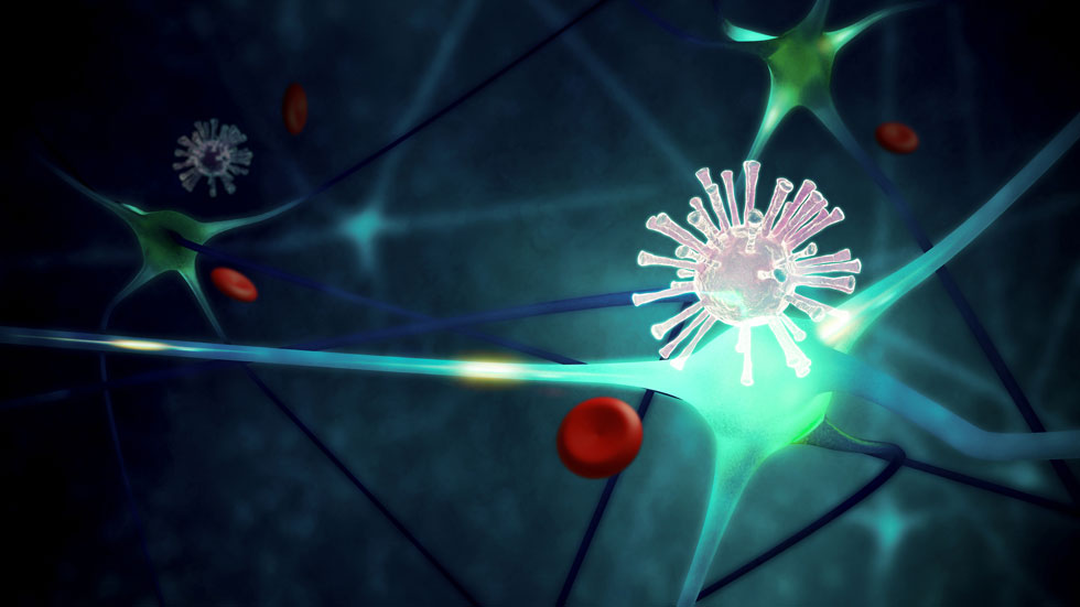
For this update, we present some pertinent updates in the literature, including new insights into COVID-19 neuropathology.
A recent publication in Brain describes the neuropathology of 9 patients who died of COVID-19 during the first wave of the pandemic with minimal respiratory symptoms, adding to our understanding of the pathophysiology by with SARS-CoV-2 leads to neurologic complications. The authors found widespread fibrinogen deposition in these brains, most prominent in the perivascular regions suggesting blood-brain-barrier disruption throughout the brain, with endothelial cell activation and platelet aggregation. Evidence of inflammation was also present throughout the brain, including evidence of complement activation and perivascular macrophage infiltration. Only rare T cells were present. Microglial clusters and neuronophagia, representing microglial activation and neuronal injury, were found, predominantly in the cerebellum and brainstem nuclei. Spatial transcriptomics analysis found dysregulation of angiogenesis-related genes in the brains of COVID-19 patients compared to controls. Despite multiple types of assays to looks for SARS-CoV-2 in the brain, including PCR and RNA-scope among others, no evidence of the virus was found in these brains. These findings suggest that SARS-CoV-2 infection may lead to immune complexes which set off a cascade of endothelial activation, platelet aggregation, and microthrombi; this in turn leads to inflammatory processes and neuronal damage in the central nervous system which may lead to the development of neurologic symptoms seen in acute COVID-19 as well as long COVID, even without direct CNS infection (Lee 2022).
Evidence of direct CNS invasion of SARS-CoV-2 is noted to be very rare, even in cases of severe neurologic COVID-19; however, several recent publications have sought further evidence of this phenomenon. A recent study published in PNAS evaluated the brain samples obtained from transethmoidal biopsy in 26 patients who had died of COVID-19. In 5 of these samples, evidence of necrosis and inflammation were found, with further evaluation for the presence of SARS-CoV-2 revealing the presence of both SARS-CoV-2 genetic material as well as the spike protein in all 5 samples. Astrocytes represented the majority of cells in which spike protein was found. The authors performed further studies, finding that SARS-CoV-2 seems to preferentially infect astrocytes in vitro in comparison to other brain cell subtypes, and that the cell receptor NRP-1 may be the basis for the virus' entry into astrocytic cells (Crunfli 2022).
A preprint of a study of 24 COVID-19 autopsy cases has also recently come to attention in the news. The authors found that these cases of patients who died of COVID-19 pneumonia displayed a variety of gross neuropathology, including cerebral atrophy, hypoxic-ischemic damage, and diffuse cerebral edema. Concurrent with other studies (including the Lee study), the authors also found platelet-rich CNS microthromboses, microglial nodules, and neuronophagia in several of the COVID-19 patients (and none in the control group). In particular, microgliosis and neuronophagia was more predominant in the brainstem of COVID-19 patients, with a correlation seen between microglial nodule density in the medulla and length of hospitalization. SARS-CoV-2 nucleic acid was found in a subset of the COVID-19 brains (10/24), and SARS-CoV-2 viral proteins were detected by immunohistochemistry in 8 cases. Of note, 5 of these subjects had defined viral protein detection within several brainstem nuclei, including the dorsal motor nucleus of the vagus, the nucleus ambiguus, the solitary tract nucleus, and the substantia nigra (Emmi 2022).
Another study showed the activation of an endogenous retrovirus, HERV-W in the brain macrophages and in other organs of patients with COVID-19. They also found HERV-W envelope protein in the serum of COVID-19 patients (Charvet et al. 2022). Based on these observations, a clinical trial is being initiated using a humanized monoclonal antibody to HERV-W in patients with Long-COVID.
Taken together, these new insights on the neuropathology of COVID-19 reflect that SARS-CoV-2 may have wide-reaching neurologic effects through a variety of mechanisms, including through endothelial activation and microthrombosis, immune-related damage, and direct CNS invasion, possibly through astrocyte infection. It also seems that the brainstem may be preferentially affected in SARS-CoV-2, whether indirectly or directly. These studies as well as further research will lead to a better understanding of the pathophysiology of the neurologic issues related to acute COVID as well as long COVID, and eventually will also hopefully lead to appropriate therapies for these entities.
References
-
Lee MH, Perl DP, Steiner J, Pasternack N, Li W, Maric D, Safavi F, Horkayne-Szakaly I, Jones R, Stram MN, Moncur JT, Hefti M, Folkerth RD, Nath A. Neurovascular injury with complement activation and inflammation in COVID-19. Brain. 2022 Jul 29;145(7):2555-2568. doi: 10.1093/brain/awac151. https://academic.oup.com/brain/article/145/7/2555/6621999 ⧉
-
Crunfli F,et al. Morphological, cellular, and molecular basis of brain infection in COVID-19 patients. Proc Natl Acad Sci U S A. 2022 Aug 30;119(35):e2200960119. doi: 10.1073/pnas.2200960119. https://www.pnas.org/doi/full/10.1073/pnas.2200960119 ⧉
-
Emmi A, Rizzo S, Barzon L, Sandre M, Carturan E, et al. COVID-19 Neuropathology: evidence for SARS-CoV-2 invasion of Human Brainstem Nuclei. MedRxiv, 2022. doi: https://doi.org/10.1101/2022.06.29.498117 ⧉ https://www.biorxiv.org/content/10.1101/2022.06.29.498117v1 ⧉
-
Charvet et al., SARS-CoV-2 induces human endogenous retrovirus type W envelope protein expression in blood lymphocytes and in tissues of COVID-19 patients. MedRxiv, 2022. https://www.medrxiv.org/content/10.1101/2022.01.18.21266111v2 ⧉
Web design by Tribal Systems