


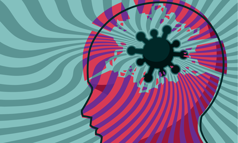
COVID-19 Update [February '23]
5 Feb 2023
By B. Jeanne Billioux, MD and Avindra Nath, MD
In the past few months, several articles have been published which provide support to the neuroinvasiveness of SARS-CoV-2, as well as its potential for viral persistence.
A notable Nature article details the in-depth evaluation of 44 autopsy cases of patients with SARS-CoV-2, including cases defined as "early" (patients having died within 14 days of illness), "mid" (died between 15-30 days of illness), and "late" (died past 30 days of illness). Autopsy tissues from widespread anatomic sites were studied systematically with digital droplet PCR (ddPCR) for viral RNA, RT-qPCR for subgenomic RNA, virus isolation with Vero E6 cells, immunohistochemistry (IHC), as well as immunofluorescence (IF), among other techniques. The authors found widespread evidence of viral RNA in a variety of tissues, including in the brain; of 10/11 brains sampled in this study, RNA was detected by ddPCR, including in a patient who died at day 230 of illness, indicating viral persistence in the brain. Moreover, the vast majority of these tissues were positive in the absence of plasma viral RNA detection, signifying that these findings were not simply due to contamination of the virus in blood. The authors also found evidence of staining of SARS-CoV-2 proteins by IHC and IF in various cells of autopsy brains, including neurons, indicating that SARS-CoV-2 does indeed have neuronal tropism. Replication-competent virus was also able to be isolated from the thalamus of one brain from a patient who died at day 13. However the amount of virus detected was very small, the number of infected cells were rare and there was no associated inflammation or pathological changes in the brain which raises questions about the significance of these findings. While this autopsy study does not address causative links between SARS-CoV-2 and Post-acute Sequelae of COVID (PASC), it does give evidence to suggest that the virus has the ability to persist in tissues outside of the respiratory tract (including the CNS) much longer than previously thought; and the role of this viral persistence in the development of PASC, requires further study (Stein 2022).
Another interesting article accepted in Neurology describes a case report of a patient with long-standing complex partial seizures who underwent epilepsy surgery 17 days after contracting COVID-19; of note, she had no new neurologic symptoms or worsening of her seizures with this viral illness. The excised brain tissue was examined for SARS CoV-2 nucleocapsid antigen by immunostaining, and extracellular vesicles were isolated from frozen brain tissue and analyzed for nucleocapsid protein by western blotting and PCR. The tissue did not stain for viral nucleocapsid antigen, but did show evidence of inflammation including infiltrates of immune cells and fibrinogen leakage into the brain parenchyma suggestive of disruption of the blood brain barrier which was not seen in controls. The extracellular vesicles from this case also were positive for SARS-CoV-2 nucleocapsid by Western blot analysis, and PCR. This case is interesting, as there was a lack of any clinical evidence to suggest a viral encephalitis in this patient; however, the unique opportunity to evaluate brain tissue proximal to COVID-19 illness revealed presence of immune activation and viral protein and RNA in extracellular vesicles. These findings suggest that the EVs may have come from the periphery, in the absence of an ongoing CNS infection. Since the patient recovered completely without any sequelae, it suggests that the detection of viral antigen alone may not be sufficient for long-term complications. This needs to be taken into consideration while investigating the pathophysiology of PASC (DeMarino 2022).
These and other cases indicate that SARS-CoV-2 may persist in the CNS without any associated pathology, and that break down of the blood brain barrier with perivascular inflammation may be caused by viral immune complexes in circulation; however, that alone may not be sufficient to explain PASC.
Due to concern that these factors may be contributing to the development of PASC, clinical trials for the treatment of PASC are being developed. One such multicenter randomized controlled trial out of Duke University is evaluating the effectiveness of Paxlovid on PASC, and is planning to start enrolling participants this month (https://clinicaltrials.gov/ct2/show/NCT05595369 ⧉). Hopefully with further ongoing studies such as those mentioned here, we will begin to untangle the puzzle of PASC and learn how to treat it effectively.
References
- Stein, S.R., Ramelli, S.C., Grazioli, A. et al. SARS-CoV-2 infection and persistence in the human body and brain at autopsy. Nature (2022). https://doi.org/10.1038/s41586-022-05542-y ⧉, https://www.nature.com/articles/s41586-022-05542-y ⧉
- Catherine DeMarino, Myoung-Hwa Lee, Maria Cowen, Joseph PSteiner, Sara Inati, Ashish H Shah, Kareem A Zaghloul, Avindra Nath. Detection of SARS-CoV-2 Nucleocapsid and Microvascular Disease in the Brain: A Case Report. Neurology Dec (2022). https://n.neurology.org/content/early/2022/12/16/WNL.0000000000201682 ⧉
- https://clinicaltrials.gov/ct2/show/NCT05595369 ⧉
Web design by Tribal Systems